Blog

Pleural Mesothelioma – Cancer of the Lung Lining

Pleural Mesothelioma is a malignant cancer in the layer of the lungs that spreading to the lungs. The spreading over the pleura created in pleural thickening. These slow down the reflexivity of the pleura and fasten the lungs in an expanding restrictive belt. With the lungs thus limited, they get inhibit in short time and a patient always run out of breath.

Pleural mesothelioma possibly:

–              carcinogenic (Diffuse and malignant)

–              non-cancerous (Localized and benign)

Non-cancerous pleural mesothelioma can be eliminated surgically, but the malignant cancers are the real haunted.

Most general between other mesothelioma circumstances, Pleural Mesothelioma is caused during exposure with blue asbestos for a long time, approximately 20 years, in the time these disease crack open fearful countenance through particular symptoms.

The symptoms of Pleural Mesothelioma

The symptoms of Pleural Mesothelioma contain hard for breathing, insomnia, aches in the chest and abdominal area, blood disgorge, laxity, weight disorder, eating disorder, lower back aches, tenacious coughing, throaty of voice, sensory disorder and trouble in swallowing.

Diagnosis of Pleural Mesothelioma

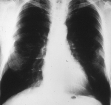
In the beginning a chest X-ray or a CT scan (computed chest tomography) was taken, which will discover a pleural thickening and an effusion. This is continuing with a bronchoscopy. Still, it has leave to a doctor for a better perceptive of the various cases. One more method is a biopsy, which can include a needle biopsy, an open biopsy, or a thoracoscopy, where a mini camera is added through the body and the tissue sample is complete for more analyze.

Treatment of Pleural Mesothelioma

The treatment is precisely equivalent with the time of the discovery of the disease, like at an early stadium the tumor can be eliminated through the surgery.

A discovering mesothelioma treatment alternative is immunotherapy, for example, Bacillus Calmette-Guerin (BCG) of intrapleural inoculation is an effective mesothelioma treatment in which an attempt to increase the immune reaction.

Radiation treatment and chemotherapy maybe the answer to the deadly pleural mesothelioma, but this only help reduce the aches, death is following with Pleural Mesothelioma.

Side effects of Treatment

The side effects of mesothelioma lung cancer treatment are damaged healthy tissues, exhaustion; too much radiation causes the skin turn into red, itchy and dry.

Next side effects of radiotherapy are diarrhea, Sickness in the stomach and vomiting, urinary discomfort and a rapid decreasing the number of white blood cells.

The typical amount life duration of someone with Pleural Mesothelioma is ended to 6 months to one year and the maximum may rise to 5 years – the magnesium-silicate mineral fibers take its toll that’s more than painful.

Other aspect  that may stimulate the chance of pleural mesothelioma are incurable lung infections, tuberculoses pleurisies, radiation (Thorotrast), vulnerability to the simian virus 40 (SV40) or mineral fibers (Zeolite) and tobacco smoking to a particular quantity.

Pleural Mesothelioma does not give someone the way for fairness. Though the life counts on many different stadiums of the malignant, it is an ultimate hazardous that draw out the life of the ordinary human.

Get a lot money? open this Ipoker domino.

Dynamite from 1940s found in Montana building during asbestos cleanup

In Montana, the cleanup crews found a decades-old dynamite in a building which has been left behind.

Lincoln County Sheriff’s Office said that there were 8 sticks of dynamite dating back to 1948. They were found in a box on the attic of the Western Montana Mental Health Center in Libby on Monday, as what reported by KRTV.

The following Tuesday, the Missoula City/County bomb-disposal team came after emergency officials cordoned off the area then they brought down the box of dynamite from the attic.

But after a while, the bomb squad reported that they found some of the deteriorated explosive came out from the box and still remained in the attic.

Libby fire and ambulance officials stand by around the building on Tuesday afternoon, the bomb squad set charges to explode and destroy the dynamite on the ground and in the attic of the building.

The official stated that the building would be demolished, it would be torn down.

Alfred Nobel, a Swedish scientist, in the late 1800s invented dynamite. It is sodium carbonate and three parts nitroglycerin to one part diatomaceous earth which can make a high explosion if perfectly mixed.

The sale of dynamite according to Nobel is to be used as an alternative way to gunpowder for a large-scale construction, for example roads and tunnel building.

But it is also a weapon in the military and revolutionaries.

You can get more interesting environment and science and space news, periodic weather updates, news and notes by following our Twitter.

High Resolution Scans are Ideal for Analyzing Asbestos Disease 

As we know that asbestos can break up into tiny fibers when they are damaged and those tiny fibers may be inhaled by human and stay in the lung to cause numerous health problems. To analyze the asbestos abnormalities in human bodies, high resolution CT scans should be used frequently. Taken by – American Journal of Roentgenology, Vol 151, Issue 5, 883-891, there is an interesting study concerned about this subject entitled, High resolution CT of benign asbestos-related diseases: clinical and radiographic correlation” by DR Aberle, G Gamsu, and CS Ray – Department of Radiological Sciences, University of California Los Angeles School of Medicine 90024. This writing is about:

An analysis about benign asbestos related pleural and parenchymal abnormalities of 100 workers who expose to asbestos in their work has been done by using high-resolution CT scans and diagnose them clinically. In the evaluation, all subjects had high resolution CT scans which is connected to conventional CT scans. High-resolution of CT scans are set as high, intermediate or low in order to evaluate for asbestosis. Different probability of basic asbestosis in multiplicity and extended observation can affect the change parenchymally. In linear regression analysis, parenchymal bands are included in the most distinctive high-resolution CT features of asbestosis along with thickened nondependent interstitial short lines. Based on the observation about  45 subjects satisfying clinical criteria of asbestosis, the probability of high-resolution CT of asbestosis was 38 (84%) for the high one, intermediate in five (11%), and low in two (4%). There are 55 subjects with 20 of them don’t have clinical asbestosis. A high probability in parenchymal abnormalities indicative was observed on high-resolution CT. A strong positive correlation between high-resolution CT and chest radiographic profusion scores for less than 0.0001, while with asbestos-related pleural thickening, it still scores less than 0.0001. In the correlation with forced vital capacity, it shows less than 0.006 and single-breath diffusing capacity scores less than 0.03 for both functional measures of restrictive interstitial lung disease. Clubbing and rales don’t have prevalence sufficiently in having statictical correlation with high-resolution CT scores. It is very sensitive to have high-resolution CT in detecting both pleural and parenchymal abnormalities in the asbestos-exposed subject. The observation od asbestos-related pleural changes are done more frequently on high-resolution CT than on conventional one or even chest radiography.

Another study about it entitled “Asbestos exposure and the risk of lung cancer in a general urban population.” By Karjalainen A, Anttila S, Vanhala E, and Vainio H – Finnish Institute of Occupational Health, Helsinki – Scand J Work Environ Health. 1994 Aug;20(4):243-50 contained that the purposes of the study is investigating lung cancer caused by asbestos based on the histological type of cancer, lobe of origin, pulmonary concentration, and type of amphibole fibers and the estimation of the etiologic fraction of asbestos for lung cancer. The methods of scanning electron microscopy is used to determine the pulmonary concentration of asbestos fibers in 113 surgically treated male lung cancer patients and 297 autopsy cases among men. Based on the pulmonary fiber concentration for all lung cancer types, squamous-cell carcinoma, and adenocarcinoma and for the lower-lobe and the upper- and middle-lobe cancers, the calculation of the age- and smoking-adjusted odds ratios of lung cancer is done.

The result of the observation is that lung cancer risk is increased based on the pulmonary concentration of asbestos fibers (f) of 1.0 to 4.99 x 10(6) f.g-1 [odds ratio (OR) 1.7] and > or = 5.0 x 10(6) f.g-1 (OR 5.3). The association od odds ratio with fiber concentrations of > or = 1.0 x 10(6) f.g-1 were higher for adenocarcinoma (OR 4.0) than for squamous-cell carcinoma (OR 1.6). The risk of asbestos-associated for lower lobe tumors was hinger than upper lobe tumors. The estimation of the rosk for anthophyllite and crocidolite-amosite fibers were similar, but not for the risk of squamous-cell carcinoma. In the greater Helsinki area, an etiologic fraction for asbestos among male surgical lung cancer patients is around 19%. The conclusion is in southern Finland, past exposure to asbestos is an essential factor for etiology of lung cancer. Pulmonary adenocarcinoma and lower-lobe tumors has a higher risk of asbestos-associated than squamous-cell carcinoma and upper-lobe tumors.

If you think that these studies are helpful, reading them in the entirety will do good for you. This article should not be used as a medical advice.

Decoration your home? Wooden Works Jepara.